- Get link
- X
- Other Apps
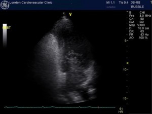
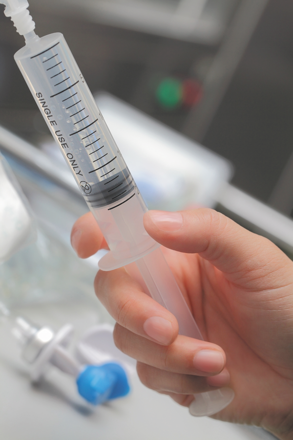
A saltwater solution called saline is mixed with a small amount of air to create tiny bubbles and then injected into your vein. A bubble study is a noninvasive test that allows physicians to assess the flow of blood through the heart.
 Andrew R Houghton How To Perform An Optimal Saline Bubble Contrast Echo Study
A bubble study is a type of echocardiogram which is the ultrasound of the heart.

Bubble study echocardiography. An echocardiogram is done to visualize the heart and its surrounding areas. Other names for this test include. The purpose of this study was to quantify the bubbles created by various quantities of agitated saline.
The Bubble Study involves a small amount of micro bubbles being sent to the heart via a vein to watch how they travel through the heart. Hepatopulmonary syndrome is an important cause of hypoxemia in children with chronic liver disease. 2142021 A bubble echocardiogram is a procedure which is designed to give a doctor an idea of how well someones heart is functioning.
This poses a potential risk for air microembolism. Bubble contrast echocardiography study for diagnosing pulmonary arteriovenous shunt in a case of hepatopulmonary syndrome Sandip Gupta Karthik Arigela Sweta Mohanty ABSTRACT Introduction. A bubble echocardiogram is typically.
112021 As such the authors review the bubble study in identifying intracardiac and extracardiac shunts including the history of its development the physics and physiology of contrast enhancement how to optimally perform and interpret an agitated saline contrast study and its safety in unique populations. The potential for misinterpretation of these bubble studies exists and therefore several false positive and false negative scenarios are illustrated and discussed. We demonstrate the clinical utility of a.
8262019 For the bubble study you will get an intravenous IV line in a vein in your arm. Hepatopulmonary syndrome was diagnosed on the basis of arterial blood gas analysis lung function testing and agitated saline contrast echocardiography in the absence of primary cardiac or pulmonary disease. You just code the echo 93306-26.
With bubble test is the accepted noninvasive standard for diagnosing PFO as it allows quantification of shunt size. In a bubble study the type of contrast used is saline or sterile salt water. We present the first attempt to identify the clinical features in patients who had cerebral.
Bubble contrast injections were performed supine or standing in a randomised order and read by a blinded observer. Better images of the heart can be produced when a contrast is used during the echocardiogram. It is typically used in conjunction with an echocardiogram in which case doctors often call it contrast echocardiography or a transcranial Doppler study TCD.
PFO patent foramen ovale. Agitated Saline Echo Agitated Saline Echo what we order at Johns Hopkins Hospital to screen for PAVMs. ASCi agitated saline contrast injection.
1212017 Such a study is labelled as negative contrast echocardiogram Fig. We describe techniques and guidelines for the detection and exclusion of a PFO. RLS right-to- left shunt.
The echocardiographic BS is frequently performed in patients that have a readily identifiable cause of stroke and whose PFO unlikely relates to the strokeTIA. 222010 A bubble study is part of the echo. 1302020 A non-invasive study typically done with an echocardiogram a bubble study helps your cardiologist to assess the blood flow and identify potential issues inside the heart.
Bubble Study A bubble study works on the principle of sound waves. Echocardiogram with Bubble Study Echocardiography is a scan that uses ultrasound sound waves to produce pictures of the heart. Our cardiologist and echo technicians do not use a contrast agent during this test.
Bubble Study findings resulted in a change in management in the minority. Figure 1 Study flow chart. The procedure is safe but the complication rate warrants informed.
A doctor will discuss a bubble echocardiogram with a patient before its performed. This fluid then circulates up to the right side of your heart and shows up on the echocardiogram image. 1 and movie clip S1.
Previous studies have recommended various safe amounts of agitated saline. 6192014 Cardiac shunts are often identified using bubble studies in echocardiography with agitated saline. Background and Purpose Detection of an intracardiac shunt is frequently sought during the evaluation of patients with cryptogenic ischemic stroke and agitated saline intravenous injection or bubble study BS is performed in most cases.
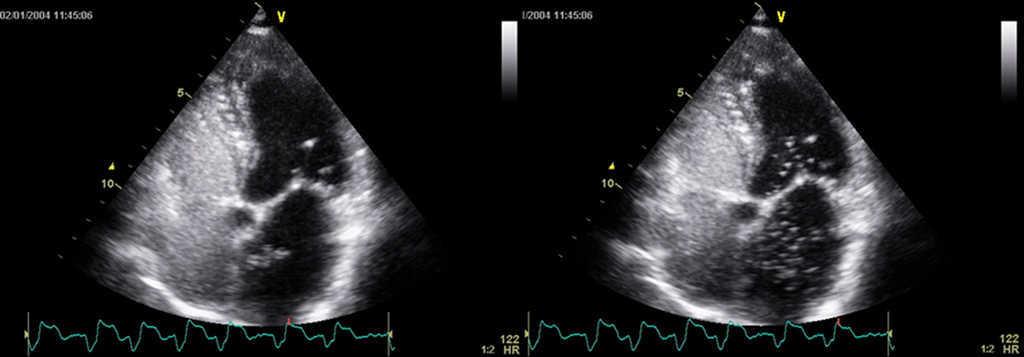
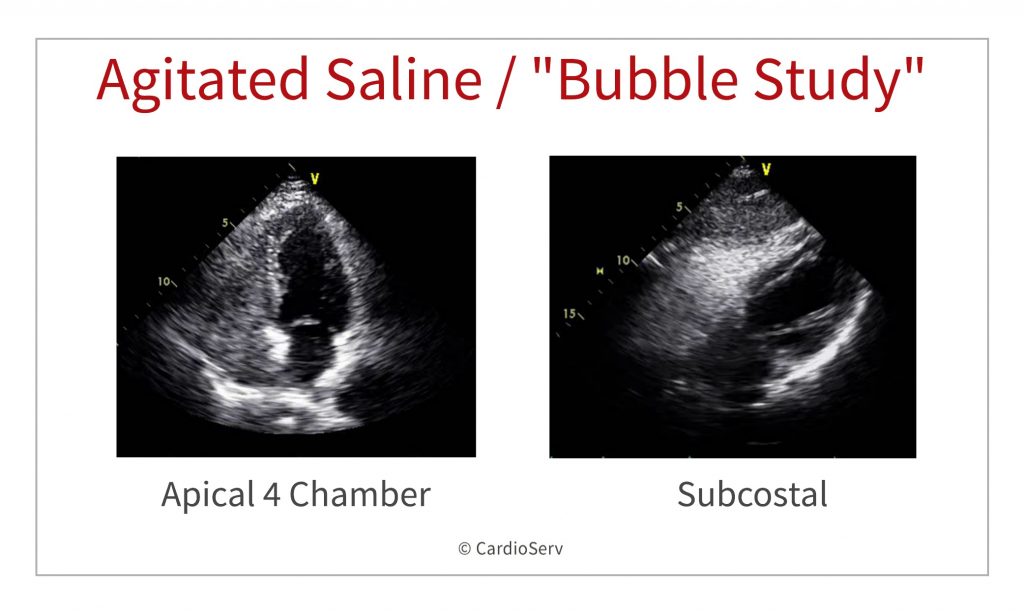
Bubble study Agitated saline transthoracic contrast echocardiograpyTTCE Contrast Echo Contrast Echocardiography. Echocardiography are currently the principal means in the diagnosis of patent foramen ovale PFO. The appearance of these microbubbles in the left atrium LA left ventricle LV or aorta commonly referred to as positive contrast echocardiogram is diagnostic of a righttoleft shunt.
It is also known as Transcranial Doppler study or contrast echocardiography.
 Transesophageal Echocardiogram Tee Bubble Study Agitated Saline Download Scientific Diagram
 Diagnosis And Quantification Of Patent Foramen Ovale Which Is The Reference Technique Simultaneous Study With Transcranial Doppler Transthoracic And Transesophageal Echocardiography Revista Espanola De Cardiologia
 7 Indications For An Echo Bubble Study
 What Is A Bubble Study Harvard Health
 Bubble Contrast Echocardiography Bubble Echocardiogram Test Llc
 Transthoracic Echocardiogram Bubble Study In A Supine Position And Download Scientific Diagram
 Echocardiogram With Bubble Study Youtube
 Anomalous Left Sided Superior Vena Cava With Cephalad Flow Ramani Gv Deible C Lopez Candales A Heart Views
 Pdf Bubbles In The Brain A Rare Complication Following Transthoracic Echocardiography
 Transesophageal Echocardiogram Bubble Study A Even Though The Right Download Scientific Diagram
 7 Indications For An Echo Bubble Study
 Saline Contrast Echocardiography In The Era Of Multimodality Imaging Importance Of Bubbling It Right Gupta 2015 Echocardiography Wiley Online Library
 A Bubble Study To Diagnose Patent Foramen Ovale Youtube

Comments
Post a Comment